


After experimenting on a hen, his dog, his goldfish, and himself, dentist William Morton was ready. On Oct. 16, 1846, he hurried to the Massachusetts General Hospital surgical theater for what would be the first successful public test of a general anesthetic.
His concoction of sulfuric ether and oil from an orange (just for the fragrance) knocked a young man unconscious while a surgeon cut a tumor from his neck. To the onlooking students and clinicians, it was like a miracle. Some alchemical reaction between the ether and the man’s brain allowed him to slip into a state akin to light sleep, to undergo what should have been a painful surgery with little discomfort, and then to return to himself with only a hazy memory of the experience.
Monitoring patients’ brains still isn’t something that medical boards require.
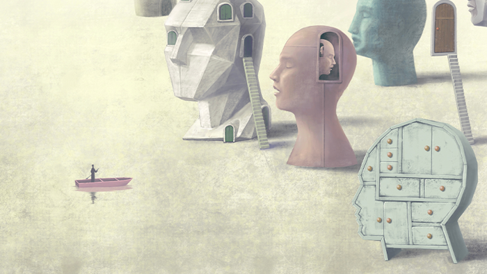
General anesthesia redefined surgery and medicine, but over a century later it still carries significant risks. Too much sedation can lead to neurocognitive disorders and may even shorten lifespan; too little can lead to traumatic and painful wakefulness during surgery. So far, scientists have learned that, generally speaking, anesthetic drugs render people unconscious by altering how parts of the brain communicate. But they still don’t fully understand why. Although anesthesia works primarily on the brain, anesthesiologists do not regularly monitor the brain when they put patients under. And it is only in the past decade that neuroscientists interested in altered states of consciousness have begun taking advantage of anesthesia as a research tool. “It’s the central irony” of anesthesiology, says George Mashour, a University of Michigan neuroanesthesiologist, whose work entails keeping patients unconscious during neurosurgery and providing appropriate pain management.
Mashour is one of a small set of clinicians and scientists trying to change that. They are increasingly bringing the tools of neuroscience into the operating room to track the brain activity of patients, and testing out anesthesia on healthy study participants. These pioneers aim to learn how to more safely anesthetize their patients, tailoring the dose to individual patients and adjusting during surgery. They also want to better understand what governs the transitions between states of consciousness and even hope to crack the code of coma.
Your brain on anesthesia
Today’s anesthetic arsenal eschews Morton’s original formula for newer, safer drugs. These include ether-based inhalants such as sevoflurane and isoflurane, and the widely used, intravenous anesthetic propofol, all of which wear off faster than early ether-based anesthetics, enabling quicker recovery. (They are also less likely to cause fires and explosions in the operating room, a regular occurrence through the first half of the 20th century.) Despite these improvements, the risks associated with excessive sedation remain high. Depending on the complexity and length of surgery, between 17 and 43 percent of patients may have cognitive problems, typically in memory and executive functions.1 These typically last only one to two weeks after surgery, but few rigorous studies have examined changes in cognitive function in the general population beyond six months after surgery. For adults over 65, the most common surgical complication is post-operative delirium, which manifests as inattention and either disorganized thought or altered levels of consciousness. Delirium can last from a few hours to several months and may be an independent risk factor for longer-term cognitive decline.2 Delirium is also preventable in 4 out of 10 patients with pre-surgery cognitive evaluations and monitoring of blood biomarkers and EEG during surgical procedures, according to the American Society of Anesthesiologists’ Perioperative Brain Health Initiative, a national nonprofit that aims to promote brain health for older adults during and after surgery.

Some studies have shown that the risk of long-term cognitive damage and post-op delirium is highest for those who already have underlying cognitive vulnerabilities, such as Alzheimer’s or mild cognitive impairment, says Paul García, associate professor of anesthesiology at Columbia University, but so far there is no consensus. The risk factors remain unclear at least in part due to varying definitions of post-operative cognitive dysfunction used in studies.
A tiny subset of patients3 (about 1 or 2 in 1,000, although estimates vary) have reported “accidental awareness,” recalling some experience of their medical procedure during general anesthesia. For many patients, researchers point out, the experience is strange but inconsequential. However, about 50 percent of those patients have reported traumas that stick with them years later and have been shown to cause long-term psychological damage and even post-traumatic stress disorder. The risk of awareness on the operating table seems to be highest when anesthesiologists need to limit anesthetic doses to protect their patients, such as when heart rate or blood pressure are already threatened following traumatic injury or emergency C-section. There are also milder and more temporary side effects of anesthesia, such as vomiting and drowsiness.
Unlike the 1846 demonstration, when Morton merely administered the anesthetic and stepped aside for the surgeon, today anesthesiology entails maintenance: Clinicians are required to monitor the patient’s vital signs, like heart rate, blood pressure, and body temperature throughout the operation, adjusting dosages as needed. But monitoring patients’ brains still isn’t something that medical boards require or that medical schools train anesthesiologists to do in the operating room.
Signal to noise
Scientists have known since the 1940s that raw electroencephalogram, or EEG, signals could be used to monitor the depth of a patient’s anesthesia-induced sleep. In the 1990s, they began to crunch this raw data to deliver a numerical index value thought to measure an anesthetized patient’s level of consciousness. But the practice of actually tracking patients’ brain activity with EEG in the operating theater is still so rare that little data is available on how widespread it is. Renowned anesthesiologist Emery Brown began using EEG to track patients’ brain waves—shorthand for electrical activity in the brain—in 2011. He estimates that over the past three years, a quarter of his fellow anesthesiologists at Massachusetts General Hospital have also begun using EEG monitoring.
EEG records the electrochemical activity between communicating neurons in the brain. As the brain lapses into anesthetic-induced unconsciousness, this activity follows a predictable pattern of sequences. Through electrodes stuck to the scalp, EEG records these sequences, which are then visualized as spiky waves on a monitor. When a patient is under general anesthesia, typically the waves crest more slowly with greater amplitude as the drugs disrupt connections between networks that keep us awake and aware. For example, propofol enhances inhibition of cortical neurons and disrupts communication across the cortex, the hub of complex functions like thinking and making sense of stimuli from the environment. It also dampens communication between the cortex, the brainstem, and the thalamus, an egg-shaped collection of neurons deep within the brain important for sensory information processing and arousal.
General anesthesia redefined surgery and medicine, but over a century later it still carries significant risks.
Brown and his colleagues have been experimenting with adjusting anesthetics according to what they see on EEG monitors. “You can actually see, based on the patterns, how unconscious someone is and you can dose your drugs accordingly,” says Brown. For example, with propofol, anesthesiologists can watch for brain waves to transition to higher amplitude, lower frequency oscillations, such as alpha and slow-delta rhythms, which signal their patient is sufficiently anesthetized and unconscious. If the patient goes too deep, the EEG becomes flatter, or slips into a potentially dangerous pattern called burst suppression, which tells the anesthesiologist to ease up on the dose. Burst suppression, indicated by a nearly flat line of suppressed activity interspersed with bursts of activity on EEG, is found in patients with inactivated brain states, such as coma or hypothermia. How easily someone slips into and out of burst suppression is influenced by age and brain health.
Not everyone is convinced that EEG monitoring will make a difference. Clinical trials testing whether EEG-guided dosing helps patients go under and recover better have so far produced mixed results,4 but more studies are underway. It’s possible that the conditions necessitating surgery and inflammation from surgery’s toll on the body could also share responsibility for any long-term effects on cognition.5
One potential obstacle to successful EEG monitoring is that the technology commonly used by those who do track brain activity relies on proprietary algorithms to process raw data and produce an index value. Some, including Brown, feel this number oversimplifies the complexity of the relationship between brain waves and the anesthesia. Due to the proprietary nature of the algorithms, it’s also hard for other scientists to evaluate the indices. The most popular EEG index tool, the Bispectral Index Monitor, was introduced in the ’90s. It uses something called bispectral processing to determine the relationship among the various EEG frequencies and then measures this value against clinical EEG and behavioral data to provide an index of a patient’s level of sedation. But it doesn’t account for the ways the brain’s response to general anesthesia changes with age,6 and so may not be as accurate in older patients.7 An index, García says, “really won’t tell you much about brain health under anesthesia.”
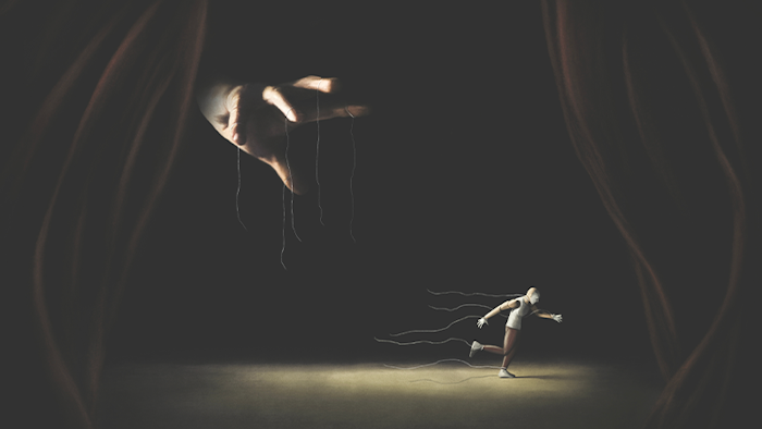
In some of these states of unconsciousness, awareness of the self and the body remains intact.
Raw EEG data may be more useful, García and others believe. Some studies published in the past decade have found links between raw EEG patterns, like burst suppression—the near-flat line of quieted neurons interrupted by spurts of neuronal chatter—and delirium or other cognitive issues post-surgery among older patients.8 In a study of 626 people ranging in age from 44 to 71, García and his colleagues saw that patients who showed burst suppression after they lost consciousness, during the maintenance phase of general anesthesia, were nearly twice as likely to experience delirium after waking up.9
Outside of the operating room, researchers have also begun conducting experiments by administering general anesthesia to healthy people who are not undergoing any kind of surgery. Studying what happens when they emerge from their anesthesia-induced sleep can provide a roadmap of how the brain should recover. Mashour and his colleagues have found that the healthy brain bounces back quickly, within just a few hours. They’ve also found that some higher, more complex functions, like executive function, are the first to return.10
Among 30 participants who underwent general anesthesia for three hours and completed cognitive tests in the following hours, abstract problem-solving skills, largely coordinated by the prefrontal cortex, appeared to recover first, while attention and reaction time recovered more slowly. Within three hours, all mental functions returned to baseline levels and the anesthetized group performed no different from the comparison group. As the participants’ cognition recovered, researchers also noticed a decrease in the frequency of a particular EEG rhythm, called the posterior dominant rhythm, which is set off when someone is resting, but awake, with their eyes closed. The researchers determined that changes in the frequency of this rhythm after general anesthesia could be a neural indicator of cognitive recovery. Though the researchers found this signal in healthy young people, it could be a potential biomarker to track recovery in surgery patients too,11 which could help anesthesiologists mitigate cognitive problems.
Getting under the hood
A better understanding of how patients wake from anesthesia and under what circumstances they struggle could also help scientists with an even knottier problem: how to treat disorders of consciousness, including coma. General anesthesia lends itself to studying these questions, as researchers can control the transition into and out of unconsciousness and track how brain activity changes as the anesthetic takes hold and wears off. Like anesthesia, coma seems to alter essential communication between different brain networks. For example, in both coma and general anesthesia, communication between the cortex and thalamus slows.
General anesthesia is “a very good tool to disentangle responsiveness and unconsciousness because you can play on the verge of these two,” says Athena Demertzi, a research associate at the University of Liège in Belgium. “It’s especially fascinating to see how these brain network configurations change in different states of anesthesia.”
Researchers are already using anesthesia to test potential coma therapies. For example, ritalin, often prescribed for ADHD, has been found to bring rats back to consciousness after general anesthesia, possibly by stimulating dopamine signaling in the brainstem’s ventral tegmental area, a node in arousal networks.12 In people with coma from traumatic brain injuries, some of those connections may remain intact. Brown and his colleagues are testing whether Ritalin could spur the recovery of consciousness in such patients via those remaining connections, and are starting a phase I clinical trial.
In her work, Demertzi wants to see if there’s a way to reach patients with consciousness disorders who may have some remaining connection to their environment, but cannot communicate. In some of these states of unconsciousness, awareness of the self and the body remains intact. One way to probe whether an unconscious person has a sense of such boundaries: Flush their ears with water.
In an upcoming study, Demertzi will give healthy volunteers general anesthesia while she monitors their brain activity via EEG and irrigates their inner ears with water. This perturbs the vestibular system, which sends information about the positioning of the body in space to the brain. In conscious people, water flooding the inner ears triggers rapid, involuntary eye movements and produces nauseating vertigo. But this reaction weakens after multiple repetitions once the brain and body habituate and learn what to expect.
Demertzi suspects that after she irrigates the ears of study subjects who have been anesthetized, they will exhibit brain activity consistent with vestibular upset, but that the response will attenuate after multiple rounds of ear irrigation. Ultimately the idea is to identify an EEG signal that tracks with this brain activity so that it could be used to determine someone’s level of consciousness.
“Using anesthesia to study the brain is not a hot topic,” but it should be, says Brown. The more we understand about what these widely used drugs do to the brain, the better we can take care of patients in all states of consciousness. 
Lead image: Agsandrew / Shutterstock
References
1. Mahanna-Gabrielli, E., et al. State of the clinical science of perioperative brain health: report from the American Society of Anesthesiologists Brain Health Initiative Summit 2018. British Journal of Anaesthesia 123, 464-478 (2019).
2. Goldberg, T.E., et al. Association of delirium with long-term cognitive decline: A meta-analysis. JAMA Neurology 77, 1373–1381 (2020).
3. Kim, M.C., Fricchione, G.L., & Akeju, O. Accidental awareness under general anaesthesia: Incidence, risk factors, and psychological management. British Journal of Anesthesia 21, 154-161 (2021).
4.Vacas, S. & Hudson, A.E. Seen and ignored: Are we undermining studies of brain health interventions before we start? Anesthesia & Analgesia 131, 464-465 (2020).
5. Azeem A., Hana, Z., Jin, Z., Suen, K.C. & Ma, D. Surgery, neuroinflammation and cognitive impairment. eBioMedicine 37, 547-556 (2018).
6. Purdon, P.L., et al. The ageing brain. British Journal of Anaesthesia 115, i46-i57 (2015).
7. Berger, M., et al. Best practices for postoperative brain health: Recommendations from the fifth international perioperative neurotoxicity working group. Anesthesia & Analgesia 127, 1406-1413 (2018).
8. Gaskell, A., et al. Modulation of frontal EEG alpha oscillations during maintenance and emergence phases of general anaesthesia to improve early neurocognitive recovery in older patients: protocol for a randomised controlled trial. Trials 22, 146 (2019).
9. Hesse, S., et al. Association of electroencephalogram trajectories during emergence from anaesthesia with delirium in the postanaesthesia care unit: an early sign of postoperative complications. British Journal of Anaesthesia 122, 622-634 (2019).
10. Mashour, G.A., et al. Recovery of consciousness and cognition after general anesthesia in humans. Elife 10, e59525 (2021).
11. Labonte, A.K., et al. The posterior dominant rhythm: an electroencephalographic biomarker for cognitive recovery after general anaesthesia. British Journal of Anaesthesia 7 (2022). Retrieved from DOI:10.1016/j.bja.2022.01.019
12. Chemali, J.J., Van Dort, C.J., Brown, E.N., & Solt, K. Active emergence from propofol general anesthesia is induced by methylphenidate. Anesthesiology116, 998–1005 (2012).
Get the Nautilus newsletter
Cutting-edge science, unraveled by the very brightest living thinkers.