


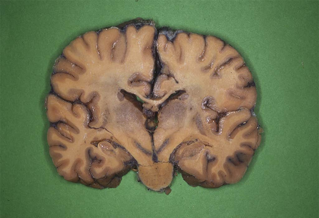
With a large blade resembling a bread knife—but without the jagged edges—Stephanie Forkel slices through the human brain lying in front of her on the dissection table. A first-year university student, Forkel is clad in an apron and protective gear. It’s her first day working in the morgue at a university hospital in Munich, Germany, where the brains of people who’ve donated their bodies to science are examined for research.
Her contact lenses feel dry because of the dense formaldehyde hanging in the air. But that’s not the only reason she squints a little harder. When she looks down at the annotated brain diagram in the textbook she’s supposed to use for reference, the real human brain in front of her looks nothing like the illustrated one.
That was Forkel’s first eureka moment: The standard reference shape of the brain and real brains were actually vastly divergent. As she continued her studies, she confirmed that, indeed, “every individual brain looked very different,” she recounts decades later.
“Every individual brain looked very different.”
A growing body of research now confirms there are plenty of physical dissimilarities between individual brains, particularly when it comes to white matter—the material nestled beneath the much-prized gray matter. And it’s not just anatomical. White matter hosts connections between the brain’s sections, like a city’s streets and avenues. So behavioral patterns can arise from even small physical differences in white matter, according to a late 2022 Science paper penned by Forkel and a colleague.1
Forkel is now one of a host of researchers probing subtle differences in white matter to better understand the extent of its role in making us who we are—including how much white matter dictates variations between people’s everyday behavior, and whether it’s implicated in how some patients recover better than others from life-threatening brain injuries.
In the past, much of what science knew about how the brain works came from case studies conducted on a small number of high-profile patients who had suffered dramatic lesions to the brain. One staple of medical lore is the case of the American railroad worker Phineas Gage who, in 1848, had a javelin-like iron rod blasted through his brain during an accident on the job. He survived—even with most of his brain’s left frontal lobe destroyed—but suffered major changes to his behavior: He became oft-irrational, his personality unrecognizable to his friends.
Cases like these ended up formalizing how neuroscientists understood what’s going on in our skulls: “this right bit” of the brain is responsible for “that bit” of cognition, and “this left bit” of the brain is responsible for “that other bit” of behavior. Over time, this modular way of thinking led to a standardized template of how all brains should look, with little room for variation.
As more sophisticated methods of studying the brain have emerged—such as MRI scans, CT scans, diffusion tensor imaging, and more—it started to become clearer this earlier view had been overly simplistic. In particular, it long overlooked the fatty and proteic—and variable—part of the brain that is white matter tissue. And new research is beginning to uncover whether and how the physical anatomy of our white matter affects how we perceive and navigate the world around us.2
For instance, there’s a chubby stretch of white matter called the uncinate fasciculus which sits between the fear-response-regulating amygdala, and the medial prefrontal cortex, which is thought to be in charge of all things logical reasoning. In some people, that portion of white matter can be a little thin and disrupted, according to Avram Holmes, a professor of psychology and psychiatry at Yale University. In these cases, the prefrontal cortex might have a harder time clamping down on the amygdala if it triggers a fight or flight response in an unnecessary situation, or vice-versa.
“You might become someone who might present as being a more anxious individual, or you could get the opposite, someone that presents as more thrill-seeking because they lose that fear,” says Holmes, whose lab specializes in looking for these types of anatomical “fingerprints” in people’s brains that correlate with behaviors.
Our white matter affects how we perceive and navigate the world around us.
“You can take information on the white matter anatomy in one group of individuals and you can use it to predict almost any behavior in a large population of folks,” Holmes says. Researchers like him have been able to spot how anatomical differences in white matter can correlate to how well we regulate our emotions,3 our ability to make decisions,4 our skill in recognizing faces,5 our ability to pay attention and be alert,6 and, to some extent, our capacity to accurately reflect7 on our own cognition and experience. Differences in white matter are also being used to try to understand differences in developing brains, such as why some adolescents develop depression8 or ADHD.9
So far, however, white matter differences only account for a very small fraction of behavioral variance between people, Holmes notes, maybe upward of only 5 percent or so. There are also aspects that researchers are better at predicting than others: IQ and working memory are easier to predict from white matter shape than, say, personality or temperament, he says. But his and others’ research continues to search for any predictive patterns emerging from the data.
There is also reason to believe that differences in people’s white matter structure affect how they age, according to Svenja Caspers, the director of the Institute of Anatomy at the University of Dusseldorf.
Not only does white matter anatomy differ from person to person to start with, but the differences are also exacerbated as time goes by. “The variability between people’s brains is increasing dramatically as we move over the lifespan into the older ages,” says Caspers, who has compared thousands of brain scans. The scientific jury is still out on the exact reason the gap between people’s anatomies grows larger over time, but it’s likely a combination of genetics, environmental factors, and lifestyle.
Differences in white matter can correlate to how well we regulate our emotions.
Population-based research already suggests that variations in white matter anatomy may line up with differences in people’s abilities as they age. For example, a 2020 study examined a group of more than 100 participants between the ages of 18 and 80 and found that anatomical differences in people’s white matter can reflect how they perform hand-coordination exercises, such as tying a shoelace or typing a message.10 Elderly individuals with healthier white matter performed slightly better in the manual experiments than those with more deteriorated, fragmented white matter. A similar study suggested the same anatomical pattern is true for how well older people perform in experiments to measure their attention capacity and executive processing, such as in the Stroop test—identifying the color of a word when the word spells out one color, but it is printed in another one, like the word “green” printed in red ink.
In general, white matter volume loss goes along with declines in processing speed, and people whose white matter shrinks more in old age also tend to have a harder time learning new things and staying focused on tasks. Some scholars believe these symptoms can be so severe as to make up a form of dementia solely related to white matter disruptions—“white matter dementia.”11 This is still just an academic concept and is not used in regular clinical practice, but it points to the powerful role for this under-appreciated anatomy we are still just beginning to understand.
New research shows that the structure of a person’s white matter might also be used someday to more accurately understand their individual trajectory after a brain injury.
For instance, zeroing in on white matter anatomy can help more precisely spot how severe a patient’s symptoms are likely to be after a stroke.12 Compared to focusing on gray matter, white matter also yields a more accurate understanding of a patient’s recovery from a brain lesion,13 according to Aaron Boes, professor of pediatrics and general neurology at the University of Iowa’s Carver College of Medicine.
This increased precision is in part due to white matter’s essential role of information conduit, so its injury can undermine functioning in gray matter sections which otherwise look fine. This function helps explain why there are sometimes patients who have a lesion in a gray area of the brain traditionally thought to be used for one type of function, like language, but have no correlated language symptoms. Or they have speech impairment symptoms but no damage on the part of the brain thought to regulate it.
Boes’ team is currently collating data from thousands of cerebral scans of people who have experienced brain injury. They are mapping white matter patterns as they correspond to outcomes after an accident: be it the lesion’s effect on cognition,14 mood, or a person’s ability to carry out difficult math tests.
It’s still too early to link any potential correlations to treatments, Boes says, but he hopes the field will continue in that direction. “There’s already a basic emergence of support for the idea that we should be individualizing treatment strategies, and targeting specific anatomical targets,” he says. Perhaps some day, a rapid image of a person’s brain post-injury—whether from a stroke or an iron rod in their brain—could help doctors understand the immediate prognosis and best treatment given that patient’s white matter presentation and the particular injury to it.
“It’s not anymore based on the theory of ‘this is generally how the brain works and therefore it should work for you, and oops, it doesn’t,’” Forkel says. “It’s really based on: ‘this is your brain anatomy.’” The same could be true some day for the diagnosis and treatment of behavioral conditions, mental health issues, and aging-related illnesses.
In the meantime, in the hopes of better understanding the role of the brain’s anatomy in cognition, Forkel now uses a more subtle tool at the morgue’s dissection table. Instead of slicing the brain into separate chunks to match the diagrams in the old-fashioned modular view, Forkel now carefully peels away at each layer until she reaches the varied and weird white matter nestled below. The question is no longer just about anatomy but now also, “What does it mean? Why are we different? Does it matter that we are different?” she notes. “There’s a huge potential to make an impact—and change the field.” 
Lead image: Jolygon / Shutterstock
References
1. de Schotten, M.T. & Forkel, S.J. Emergent properties of the connected brain. Science 378, 505-510 (2022).
2. Johansen-Berg, H. Behavioral relevance of variation in white matter microstructure. Current Opinion in Neurology 23, 351-358 (2010).
3. Pedersen, W.S., et al. Individual variation in white matter microstructure is related to better recovery from negative stimuli. Emotion 22, 244-257 (2022).
4. Boorman, E.D., O’Shea, J., Sebastian, C., Rushworth, M.F.S., & Johansen-Berg, H. Individual differences in white-matter microstructure reflect variation in functional connectivity during choice. Current Biology 17, 1426-1431 (2007).
5. Unger, A., Alm, K.H., Collins, J.A., O’Leary, J.M., & Olson, I.R. Variation in white matter connectivity predicts the ability to remember faces and discriminate their emotions. Journal of the International Neuropsychological Society 22, 180-190 (2016).
6. Niogi, S., Mukherjee, P., Ghajar, J., & McCandliss, B.D. Individual differences in distinct components of attraction are linked to anatomical variations in distinct white matter tracts. Frontiers in Neuroanatomy 4 (2010).
7. Baird, B., Cieslak, M., Smallwood, J., Grafton, S.T., & Schooler, J.W. Regional white matter variation associated with domain-specific metacognitive accuracy. Journal of Cognitive Neuroscience 27, 440-452 (2015).
8. Vulser, H., et al. Early variations in white matter microstructure and depression outcome in adolescents with subthreshold depression. The American Journal of Psychiatry 175, 1255-1264 (2018).
9. Nagel, B.J., et al. Altered white matter microstructure in children with attention-deficit/hyperactivity disorder. Journal of American Academy of Child & Adolescent Psychiatry 50 283-292 (2011).
10. Adab, H.Z., et al. Fiber-specific variations in anterior transcallosal white matter structure contribute to age-related differences in motor performance. NeuroImage 209, 116530 (2020).
11. Filley, C.M., Franklin, G.M., Heaton, R.K., & Rosenberg, N.L. White matter dementia. Neuropsychiatry, Neuropsychology, and Behavioral Neurology 1, 239-254 (1988).
12. Forkel, S.J., et al. Anatomical evidence of an indirect pathway for word repetition. Neurology 94 e594-e606 (2020).
13. Reber, J., Hwang, K., Bowren, M., & Boes, A.D. Cognitive impairment after focal brain lesions is better predicted by damage to structural than functional network hubs. Proceedings of the National Academy of Sciences 118, e2018784118 (2021).
14. Bowren, M., et al. Multivariate lesion-behavior mapping of general cognitive ability and its psychometric constituents. The Journal of Neuroscience 40, 8924-8937 (2020).
Get the Nautilus newsletter
Cutting-edge science, unraveled by the very brightest living thinkers.