
People say it looks like broccoli, a tree, a bowl, a pizza, a crescent moon, a Frisbee, a pancake. Etymologically, “pancake” is closest: Placenta means cake in Latin and comes from the Greek word plakoenta, which means flat cake. Per a Greco-Roman recipe that’s been dated to 160 B.C., in order to make the round, flat dessert called “placenta,” you need thin sheets of dough, cheese, honey, and bay leaves. If you’re trying to make the organ called “placenta,” the recipe is, of course, trickier than that.
Ancient civilizations gave the placenta great symbolic and spiritual importance. In some cultures, it was considered sacred because it protected the baby and represented the original mother-child bond. People often buried or cremated the organ in rituals that persist in some cultures today: For example, members of the Southeast Asian Hmong community bury the placenta at home, under the belief that when a Hmong person dies, their soul returns to its birthplace and reunites with the placenta, which helps it travel to the spirit world.
Most contemporary societies treat the placenta as biological waste and toss it straight into the incinerator after birth. But increasingly, the organ is ending up on the lab table, where it can teach us how it functions as an endocrine and immune interface between mother and child.
Research into placental formation has illuminated the underlying mechanisms of many pregnancy-related pathologies. Most notably, it has helped the medical community gain a greater understanding of preeclampsia, one of the most common pregnancy disorders, affecting approximately 5 percent of pregnant women.
Clinically, preeclampsia is characterized by high blood pressure and elevated levels of protein in the urine (proteinuria). In serious cases, it can provoke multi-organ lesions and hemolysis, elevated liver enzymes, and a low platelet count (HELLP syndrome). Preeclampsia is a multifactorial disorder involving genetic, immune, and vascular factors in both the mother and the fetus. One school of thought, though not the only one, suggests that these factors compromise the placenta’s capacity to adequately develop the blood vessels that connect it to the mother’s circulatory system. As a result, those vessels can become narrower than normal, leading to dysfunction.
At the start of pregnancy, when the fetus is small and needs little oxygen and nutrition, reduced blood flow is not a major problem. But as the pregnancy progresses and the fetus grows, so do its demands. When that happens, the placenta compensates for this limited blood flow by signaling the mother’s body to send more blood — but the supply remains insufficient, and the placenta tries again. This creates a vicious cycle that produces preeclampsia symptoms: hypertension, swelling, headaches, vision problems, and more.
Happily, studying the placenta can contribute to biomedical science far beyond our understanding of preeclampsia — and even beyond maternity itself. In fact, it may also hold clues to solving perhaps one of the most consequential medical mysteries: how cancer cells reproduce.
To be clear, “cancer” refers to a group of diseases characterized by the growth of abnormal cells that divide rapidly and uncontrollably. These cells can invade healthy tissue and form new blood vessels to supply themselves while avoiding the immune system’s soldiers. This is true of placental cells, too. Its cells can divide rapidly, invading the mother’s endometrium, forming new blood vessels to supply the baby, and eluding the mother’s immune system.
This strange parallel has stirred up considerable interest among scientists. A major line of investigation in cancer research now uses placental cells to better understand cancer biology and develop new treatments. It’s worth emphasizing here that placental cells are not cancerous — not at all.
Rather, they are essential for a fetus to develop and survive, growing in a precise, coordinated, and controlled way. Cancer cells, by contrast, multiply uncontrollably and damage the organism. Their objective is nothing like that of placental cells; they simply use similar strategies. As a result, international initiatives like the NIH Human Placenta Project have emerged to study how the placenta affects maternal and fetal health not just during pregnancy but throughout the lives of the mother and child.
Common medical wisdom is that organ development begins with the heart. But technically speaking, that is not quite true. A baby’s first organ to develop is not its heart or kidneys or brain but its placenta, which begins forming six to 10 days after fertilization and is up and running by the 12th week.
Nearly all of its cells contain the baby’s DNA, though a handful contain the mother’s. Most come from the endometrium, the inner layer of the uterus, called the decidua, during pregnancy. Both the baby and the placenta develop from the cell formed when the egg and sperm fuse. The placenta’s origin, then, is fetal.
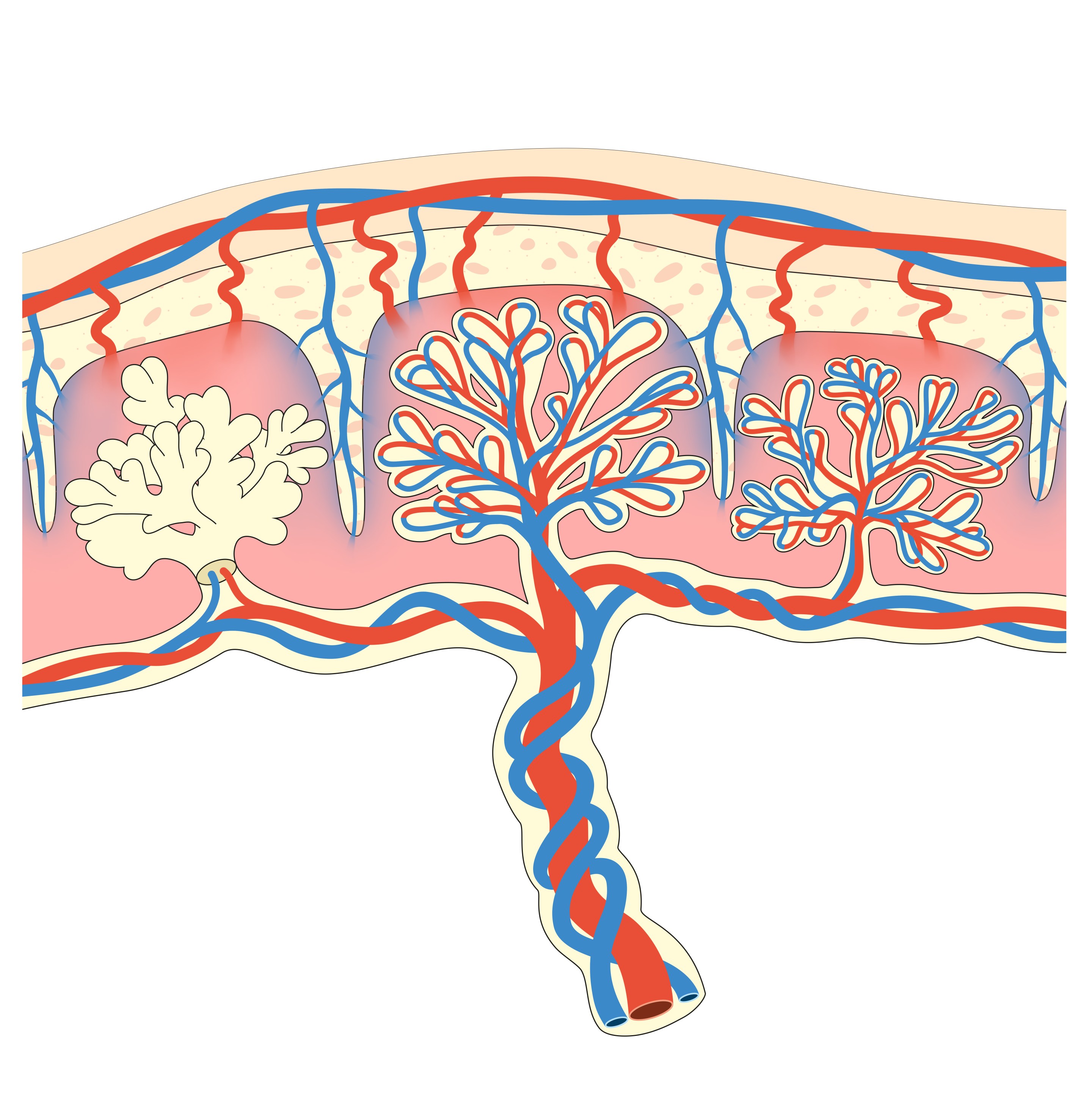
From there, the placenta has many duties. Its best-known role is mediating between maternal and fetal bloodstreams. It is no mere bouncer deciding what can or cannot pass; it is a sophisticated entity charged with numerous tasks key to the pregnancy’s success.
Until the baby can manage on its own, the placenta is its primary caregiver. It takes on the roles of the lungs, gut, liver, and kidneys, since the fetus cannot yet breathe, digest, or clear its own waste. The placenta draws oxygen and nutrients from the mother’s blood, transforms many of them into forms the baby can use, and passes them into the fetal circulation, while carbon dioxide and waste products travel back the other way, to be cleared by the mother’s lungs and kidneys. The heart is the one organ the baby runs itself from very early on: it starts beating around three weeks after fertilization, and it is the fetal heart — not the placenta — that drives blood through those exchange surfaces.
The placenta does not just serve as an interface. It is also the pregnancy’s control center. It generates and releases substances like hormones and cytokines that travel through the bloodstream and modify the behavior of the mother’s other organs.
For instance, the placenta produces human chorionic gonadotropin (hCG), which maintains endometrial receptivity, allowing a zygote to implant. (Pregnancy tests check whether hCG is present.) After fertilization, the cells that will become the placenta produce hCG to prevent the corpus luteum — a temporary, hormone-generating organ — from disintegrating. If it did, it would no longer produce progesterone, which is necessary to maintain the endometrium in ideal conditions for implantation. hCG also prevents the corpus luteum from degenerating, which would otherwise cause menstruation. As the pregnancy progresses, the placenta no longer needs the corpus luteum, since it can produce its own progesterone and keep the endometrium intact.
The placenta also secretes various estrogens and placental lactogen, which stimulate the development of mammary tissue, and produces hormones such as relaxin, which increases mobility in joints and ligaments. Placental hormones also fine-tune the cardiovascular, metabolic, and immune systems, among others, to meet the demands of each stage of pregnancy. This marvelous organ even makes the inflammatory cytokines and prostaglandins that help start labor.
These substances help maintain a stable pregnancy by enabling the mother’s body to adapt dynamically to changing metabolic, immune, cardiovascular, and neurological demands. Research I’ve conducted in my lab indicates that these hormones also modify the mother’s brain to optimize gestation, parturition, and the postpartum period. And perhaps, hidden within this still-mysterious dialogue between the placenta and the brain lie some of the clues we need to better understand and protect maternal mental health. 

This story is reprinted with permission from MIT Press Reader. It is adapted from A Mother’s Brain.
Lead image: Aldona / Adobe Stock