
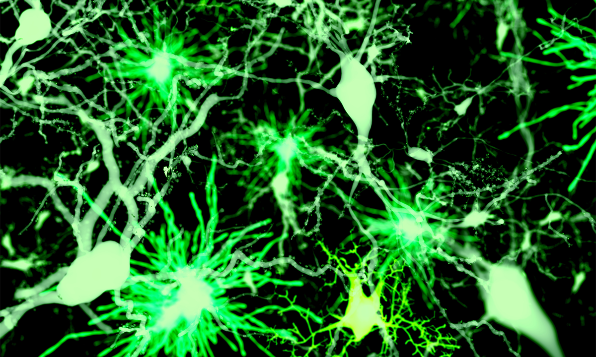
One of the ongoing challenges in understanding the workings of the human brain is being able to visualize its activity in real time. Our brains are collections of nerve cells, or neurons, that receive, transform, and send signals to other cells to create thoughts, decisions, and memories. To date, researchers have illuminated the outgoing signals from nerve cells using techniques like electrophysiology but have found the incoming signals too fast and faint to capture.
Now, described in a recent paper published in Nature Methods, neuroscientists have devised a way to detect incoming chemical signals. “What we have invented here is a way of measuring information that comes into neurons from different sources, and that’s been a critical part missing from neuroscience research,” explained lead study author Kaspar Podgorski at the Allen Institute in Seattle in a statement.
Podgorski and an international team of collaborators from the United States, Germany, Italy, London, and Austria engineered variants of a protein—iGluSnFR—to record incoming brain cell signals. Nerve cell signals bridge the gap between neurons by sending chemical messengers across the gap, or synapse. The most common messenger for learning, memory, and feelings is the molecule glutamate, for which the iGluSnFR protein is a good tracking indicator.
Read more: “A New Doorway to the Brain”
By testing the performance of 70 variants of iGluSnFR in mouse brains, the researchers discovered two variants that were sensitive enough to detect even the faintest incoming neural signals. The glutamate indicators were tested in mouse models in various regions of the brain—including the neocortex, thalamus and hippocampus, and midbrain—and proved able to provide a window into information flow between neurons of various types. Coupled with existing techniques to monitor outgoing signals, the iGluSnFR variants offer a way to interpret entire through-puts of information in the brain.
“I feel like what we’re doing here is adding the connections between those neurons and by doing that, we now understand the order of the words on the pages, and what they mean,” Podgorski continued.
The scientific advance has implications for treating a range of diseases that are linked to disruptions in glutamate signaling, including Alzheimer’s, schizophrenia, and autism. Being able to visualize the activity of synapses paves the way for understanding the mechanisms underlying brain disorders and then developing drugs that restore normal synaptic function. 
Enjoying Nautilus? Subscribe to our free newsletter.
Lead image: Juan Gaertner / Shutterstock