The Nikon Small World photography contest doesn’t celebrate weather, wildlife, or natural landscapes. Instead, it’s meant to honor images of the world at the smallest possible scales, offering unique perspectives on the details of the environments around us.
This year, judges cited 76 of the 1,803 entries as either an award winner, runner-up, honorable mention, or image of distinction.
The contest’s mission is to “showcase the beauty and complexity of things seen through the microscope,” and this year’s winners definitely fulfilled that promise. There was everything from inkjet printers and pens, to wasps and neurons—all in high resolution and ultra close-up (but of course).
German Ole Bielfeldt won first place for his shot of the inner workings of an inkjet printer. Getting the final photograph involved a custom printing rig and a custom microscopic camera rig, using a 20x magnification. Credit: Nikon Small World
Second place went to John-Oliver Dum, also from Germany, who used a 20x magnification to grab this image of a jumping spider (Salticus scenicus) with particles of pollen on it. Credit: Nikon Small WorldFor the third-place photo, Kaelin Wulf and Rachel Flannery employed 5x magnification to show neurons in an expanded mouse brain. Both work at the Allen Institute in Seattle. Credit: Nikon Small WorldI bet you can’t you guess what this is: the very tip of a ballpoint pen, snapped at 20x magnification by Justin Theodore Benttinen from the U.S. Credit: Nikon Small WorldThe inner structure of a closed daisy, captured at 10x magnification by Michael Crutchley of the U.K., using a darkfield microscopic photography technique to foreground the flower. Credit: Nikon Small World
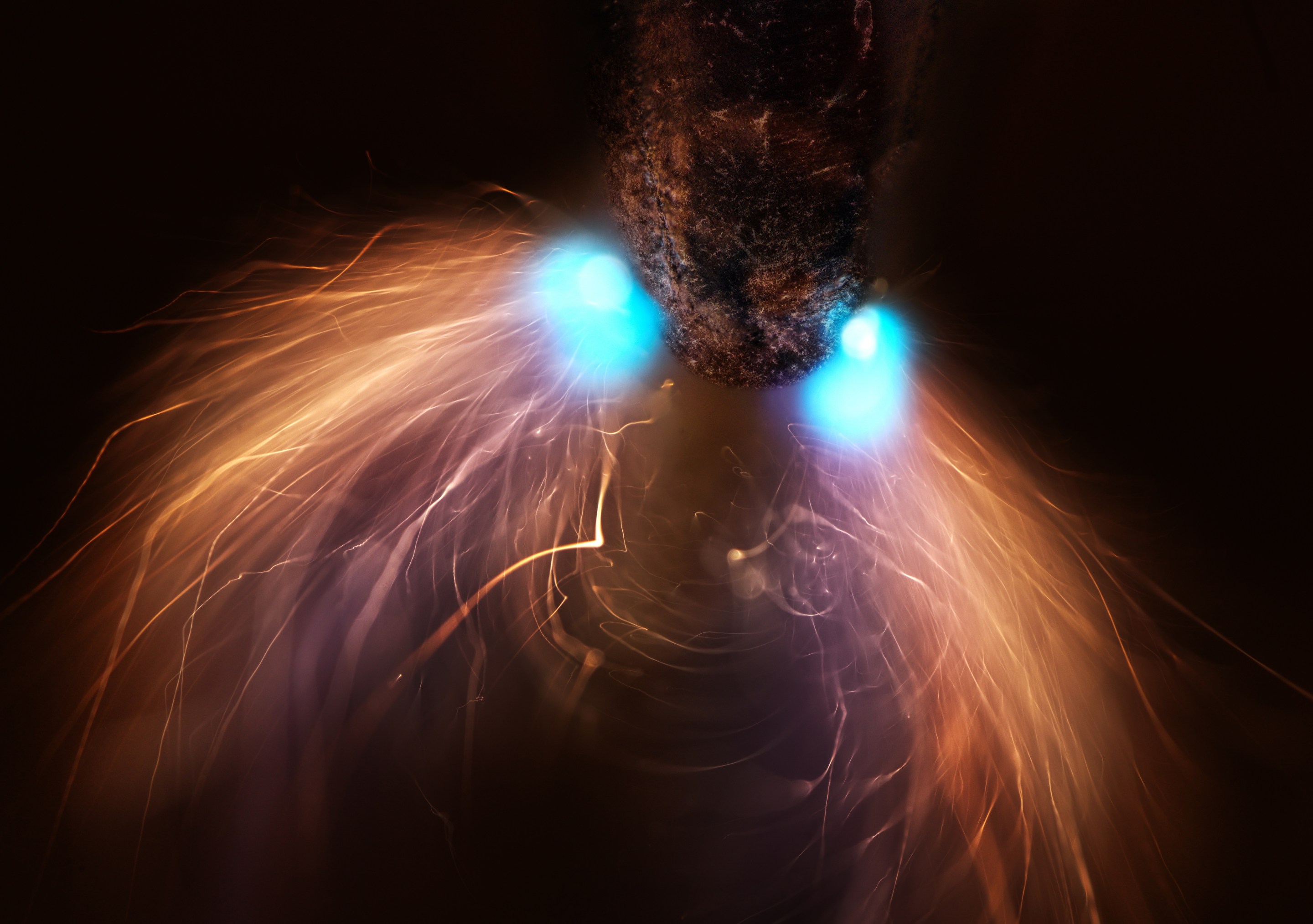
Another one where it might take you a minute to figure out what you’re looking at: a Hemitrichia slime mold spore structure, taken at a 10x magnification, by Eric Epstein of the U.S. Credit: Nikon Small WorldThese are human neurons reprogrammed from skin cells, growing in a microfluidic chamber. The image, photographed at 20x magnification, is the work of Dr. Bruno Cisterna and Dr. Eric Vitriol, from Augusta University in Georgia. Credit: Nikon Small WorldIt looks like some sort of alien, but this is actually pencil lead (at the top) being exposed to a high-voltage electrical discharge. The photograph, at 10x magnification, is by Dr. Marcel Clemens from Italy. Credit: Nikon Small WorldCourtesy of Dr. Francisco Lázaro-Diéguez from the Albert Einstein College of Medicine in New York, this is a 40x-magnification image of a dedifferentiated liver endothelial cell from the lining of a blood vessel. Credit: Nikon Small World
A parasitoid wasp (Conura sp.) pulling itself out of a butterfly chrysalis (Opsiphanes sp.) after using it for food, as shot by Michael Parra Puentes from Colombia at 3.7x magnification. Credit: Nikon Small WorldA ghostly image of a 20-day-old sea urchin larva, lit up by blue fluorescent nuclei, taken at 20x magnification. The photographer behind it is Svenja Carolin Kling, working for the Scripps Institution of Oceanography. Credit: Nikon Small World
David Nield has written about science and technology for more than 20 years. His work has appeared in a wide range of publications, including Wired, Popular Science, The Guardian, and Gizmodo.