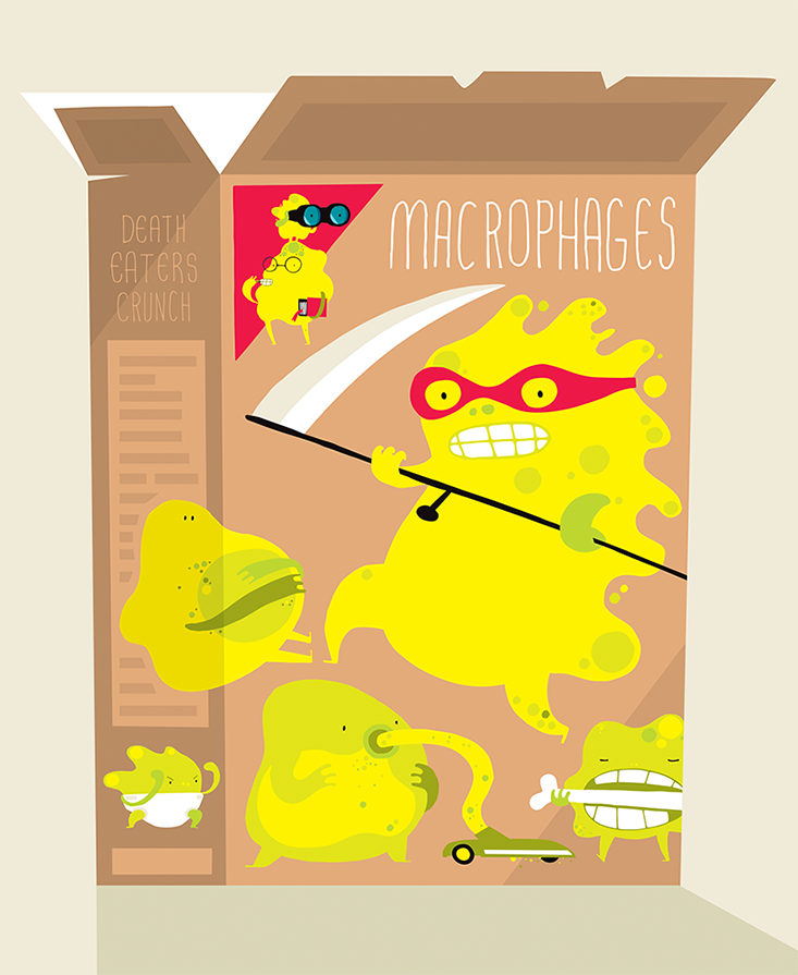
The word “macrophage” means “a big eater” in Greek, and it’s a fitting description. Macrophages are our body’s garbage-devouring cells. They consume the pathogens in our bloodstream, the necrotic cells from our flesh and bones, and the dust in our lungs. Present in every part of our body, they’re a key part of our immune system.
Scientific illustrations of macrophages look like little fried eggs, but such images belie the sophistication and variety of these cells. From their microscopic centers, macrophages extend chemical, physical, and mechanical sensors which can detect cellular changes such as illness and death. Based on what they find, macrophages can secrete hundreds of proteins to trigger the immune system’s defense against pathogens, the growth of new bone cells, and even the termination of a pregnancy.
Scientists don’t have a complete enumerated portrait of all the variations of macrophages that take out our body’s trash, but we selected several types to show their vital role in our health and reproduction.
Sinusoidal macrophages:
the scouts and the ground troops
Sinusoidal macrophages are both scouts and the first line of defense against pathogens in our body—they reside in the spleen, which filters bacteria and viruses from the blood stream. In the battle against infections, the human immune system depends heavily on lymphocytes called T-cells and B-cells. These cells are very good at defending our body against infectious agents, but not at identifying them. Sinusoidal macrophages identify pathogens, assess their danger level, and summon the T- and B-cells for an attack.
Through a process called phagocytosis, macrophages engulf the pathogen and assault it with enzymes that break it down into its constituent parts. Yung Chang, a professor at the Biodesign Institute at Arizona State University, likens them to ground troops.
“Macrophages are trying to fight a war and when you mobilize your army, you use all parts of your arsenal to destroy the enemy,” she says. And when the war is over, macrophages swoop in to take out the bodies.
Microglia: the brain matter recyclers
Microglia are macrophages that reside in the brain and central nervous system; they respond to inflammations and infections of the brain and spinal cord. The blood-brain barrier protects the central nervous system from most pathogens, but viruses such as herpes and HIV can breach the barrier and infect the brain and nerves. In other parts of the body, macrophages summon T- and B-cells, but in the brain and spinal cord microglia have no backup and have to defend against pathogens on their own.
Microglia also devour damaged neurons and plaques. Microglia also transform some of their digested waste into nutrients that they feed back to neurons. “From a waste point of view, some of the material can be recycled and reused,” says Chang, adding that microglia secrete the growth factors which “help neurons grow.”
Researchers have an ongoing debate about whether microglia mitigate or aggravate neurodegeneration. Studies have found that microglia can destroy the amyloid beta protein implicated in formation of damaging Alzheimer’s plaques. At the same time, microglia can secrete neurotoxins that cause inflammation, which can kill neurons.
Alveolar macrophages: the lung dusters
Known as dust cells, alveolar macrophages live in the lungs, preventing dust particles from accumulating as they do under the couch. Alveolar macrophages have a sophisticated sensor system that detects foreign particles among the lungs’ native cells. For an alveolar macrophage, the sharp edge of a speck of dust is a cue to start eating. “If the edge is more sharp, or has curvature, they’re likely to engulf,” says Chang.
“If the particles have a smooth surface, the macrophages are less likely to eat.”
Through phagocytosis, alveolar macrophages can digest many kinds of dust, but not all. If an alveolar macrophage absorbs a particle it can’t break down, the cell itself becomes an irritant to its host organism, and either dies or gets expelled through coughing; such malfunctioning macrophages can cause inflammations.
Osteoclasts: the bone rebuilders
Osteoclasts are macrophages that help regenerate our bodies’ bones. Over a seven-year cycle, the cells that make up the bones of the human skeleton die off and have to be replaced. Osteoclasts identify, absorb, and digest dead and dying bone cells, clearing space for osteoblasts—the bone building cells—to create new growth. This cycle explains how bones can lengthen and change shape as a person grows and why a shattered bone can heal.
Osteoclasts and osteoblasts work in tandem; an imbalance can cause osteoporosis—a medical condition in which bones become fragile and brittle, and break easily.
Decidual macrophages: the pregnancy guardians
Decidual macrophages, which live and work within the lining of the uterus, are the first cells to arrive at the implantation site of the embryo. “Implantation is like making a wound in the uterus,” says Gil Mor, Division Director of Reproductive Sciences at the Yale School of Medicine. “Implantation starts breaking the cellular epithelium in the uterus.” Cells start dying and decidual macrophages arrive to clean up.
But instead of summoning T- and B-cells, decidual macrophages send messages to the mother’s immune system directing it not to reject the embryo. As long as the fetus is growing normally, decidual macrophages “oversee” its development throughout the pregnancy. But if the embryo develops genetic anomalies, decidual macrophages could force pregnancy termination by interfering with the growth of trophoblasts—the protective outer layer of embryo cells. “If something goes wrong, the macrophages will change their phenotype and instead of guiding, they will form a barrier to stop the trophoblast’s progress,” says Mor.
But such a response is not always warranted, Mor says. An infection in the placenta can create proteins that change the behavior of macrophages from fetal guardian to fetal attacker, and then macrophages can cause a miscarriage or send the mother into unwanted pre-term labor. Mor seeks ways to either prevent such proteins from reaching macrophages, or to alter their response.
Hofbauer cells: the fetal protectors
While decidual macrophages work on the mother’s side of the placenta, Hofbauer cells work on the fetal side. Through phagocytosis, Hofbauer cells absorb invaders, such as HIV and herpes viruses, and deploy enzymes that break them down before they can transfer from mother to fetus. But even when an infection doesn’t penetrate the placental barrier, messenger proteins associated with it can sometimes cross over. Hofbauer cells react to these proteins like a real infection on the fetal side.
This can cause a condition called fetal inflammatory response syndrome, in which the fetal immune system is activated despite the absence of an infectious agent, which can lead to developmental defects in the fetus. Outside of this rare condition, Hofbauer cells appear to be fundamental to life from the very start—they have been linked to building the first blood vessels in the growing fetus.
Tumor Macrophages: the ultimate death-eaters
When a malignant formation is treated with radiation or chemotherapy, special tumor-associated macrophages gather up to devour the killed cancerous cells. Scientists experiment with tumor-associated macrophages to enhance the effects of traditional cancer therapies.
In advance of chemotherapy or radiation, scientists may be able extract tumor macrophages from the patient and make them grow cancer-killing viruses within their cellular bodies. The “armed” macrophages would be then reinserted into the patient to play a double role. First, they’d penetrate deep into the treated tumor to release thousands of copies of the virus. Then, macrophages would devour the tumor cells killed by the chemotherapy, while the virus would destroy the cells that managed to survive it.
Claire Lewis, a professor at the University of Sheffield Medical School, runs such experiments on mice, hoping to soon offer the therapy to humans. She arms macrophages by inserting an oncolytic virus, which specifically infects and kills cancer cells, and a fragment of DNA called a plasmid, which acts as a switch. When tumor cells grow they deplete oxygen around them, a condition tumor-related macrophages can detect and are naturally drawn to. The low oxygen levels also activate the plasmid switch which causes viruses inside macrophages to replicate. Macrophages eventually explode, and viruses infect and destroy cancer cells.
Patchen Barss is a Toronto-based journalist specializing in new ideas in science and the humanities. He is the author of The Erotic Engine: How Pornography Has Powered Mass Communication from Gutenberg to Google, and has written for many publications, including Saturday Night, the National Post and the Montreal Gazette.


