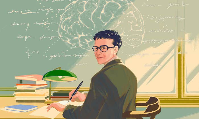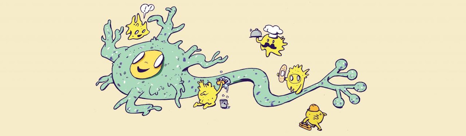

When we speak of brain cells we usually mean neurons: those gregarious, energetic darlings of cell biology that intertwine their many branches in complex webs and constantly crackle with their own electric chatter. But neurons make up only half the cells in the brain. The rest, known as neuroglia or simply glia, have long lived in the neuron’s shadow.
French physiologist Henri Dutrochet first documented glia in 1824, though he had no idea what they were—he simply noted globules between the nerves of mollusks. In 1856, German biologist Rudolf Virchow gave those blobs the name “neuroglia,” describing them as “a sort of putty in which the nervous elements are embedded.” In the following decades, scientists learned that this putty was in fact made of individual cells—at least six major types, we now know—that formed intricate structural networks with both neurons and blood vessels. Yet they still regarded glia (which is Greek for “glue”) as mere fluff ‘n stuff, the brain’s packing peanuts, an inert plasma holding everything else in place.
By the early 1900s, that notion had begun to erode. Many leading neuroscientists proposed that glia were in fact much more active than previously realized: Perhaps they were feeding neurons, or helping them communicate, or repairing them after injury. From the 1960s onward, thanks in large part to a suite of sophisticated laboratory tools, neuroscientists confirmed that glia are the brain’s architects, doctors, police, janitors, and gardeners. In the last five years, researchers have finally brought glia into the limelight as the highly dynamic, incomparably versatile, and indispensable partners of the neuron. Here are five recently discovered roles glia play in the brain:
Wiring
Neurons are not always born where they are meant to reside. In the developing brain, so-called radial glial cells form a widespread lattice of cables along which neurons crawl like inchworms to their permanent homes. When this scaffolding is no longer required, radial glia transform into other kinds of glia, such as starburst-shaped astrocytes and octopus-like oligodentrocytes, or even into neurons. Scientists recently discovered that a specific subset of radial glial cells are fated to become neurons in the upper-most region of the cerebral cortex, the brain’s wrinkly outer layer responsible for our most sophisticated mental talents.
Because the cerebral cortex of the human brain is so large and dense for a mammal our size, these glial cells likely played a key role in our evolution. A series of studies in the last three years have also confirmed that some glial cells excrete molecules that promote the formation of new connections between neurons, while others engulf and digest weak and underused synapses, changing the brain’s micro-circuitry throughout life.
Clearing Clutter
Every organ in the body needs a clean-up crew: a means of clearing away superfluous fluid, dead cells, and lingering cellular debris that could impede business as usual. The brain is no exception. Scientists have known for years that finely branched glial cells called microglia play a major role on the brain’s waste management team. Microglia roam about scavenging harmful tangles of proteins, the remains of dead cells, and bits of unneeded DNA. But a study published just last year indicated that microglia are essential for eliminating clumps of amyloid beta and other protein clusters associated with Alzheimer’s and related neurodegenerative disorders.
Microglia are not the only members of the glia tribe that help take out the trash, though. Three years ago, Jeffrey Iliff, then of the University of Rochester Medical Center, and his colleagues injected fluorescent molecules into the fluid surrounding the brains of live mice. The molecules traveled through a previously unrecognized network of channels formed by glia known as astrocytes, which flank arteries and veins. Perhaps these glial ducts, Iliff and his team surmised, act as a drainage system for the brain. When they introduced amyloid beta into the rodents’ brains it was indeed cleansed away via the astrocyte aqueduct.
Helping Neurons Talk
The oligodendrocyte precursor cell (OPC) is one of the most unique and active types of glia. OPCs eventually mature into adult oligodendrocytes that wrap their many tentacles around neuronal branches, sheathing them like rubber insulating electrical wire. Scientists discovered more than a decade ago that OPCs form synapses with neurons and change their own behavior based on the electrical signals they receive from those neurons. They are the only glial cells to do this. Now, emerging evidence indicates that the communication between OPCs and neurons goes both ways.
The surfaces of OPCs are studded with a distinct protein known as NG2, and in a study published last fall, Dominik Sakry and Angela Neitz of Johannes Gutenberg University Mainz showed that the electrical impulses OPCs receive from neurons sometimes trigger enzymes to cleave NG2 from the cell membrane, allowing the protein to drift away and contact nearby neurons. When the cleaved fragments of NG2 bind to neurons, they make the cells more responsive to neurotransmitters such as glutamate, which are essential players in neuronal communication. When Sakry and Neitz eliminated either NG2 or its associated enzymes from mice, the animals’ ability to pick up sensory information was impaired: They were slower than typical mice to realize that a repeated startling sound was innocuous, and they showed less interest in new smells. That suggests the cross-talk between neurons and OPCs is not mere idle chatter, but rather an essential dialogue that underlies behavior.
Helping You Breathe
The glia known as astrocytes wrap tightly around blood vessels feeding neurons, which puts them in an excellent position to monitor blood contents and adjust circulation as needed. Alexander Gourine of University College London and his colleagues studied how astrocytes in a rat brain might respond to fluctuating blood levels of oxygen and carbon dioxide. First, they genetically engineered the astrocytes of living rats to glow when the cells revved up their internal calcium signals, which help orchestrate activities within the cell. Then they exposed the astrocytes to differing pH levels.
Only astrocytes in the medulla oblongata, a part of the brain stem that controls breathing and heart rate, responded. When those astrocytes detected a drop in blood pH, which would correspond to elevated levels of carbon dioxide, they increased their internal calcium signaling and began to secrete adenosine triphosphate (ATP)—a molecule used to store energy and perform a wide range of cellular tasks. The ATP stimulated surrounding neurons to fire, which increased the breathing rate in live rats, eventually bringing more oxygen to the brain. Raising the pH, which would correlate with oxygenated blood, had the opposite effect. This suggests that glia are crucial for every breath you take.
Making You Smart
In Daniel Keyes’s 1958 short story Flowers for Algernon, scientists perform experimental brain surgery on a man named Charlie Gordon to dramatically increase his intelligence. First, though, as so often happens in medical research, they test out the procedure on a mouse—the eponymous Algernon. A few years ago, scientists did something spookily similar (to a mouse, that is, not a human). Steven Goldman and Maiken Nedergaard of the University of Rochester Medical Center and their colleagues injected immature human brain cells into the heads of infant mice.
A few months later, those part-human mice performed much better on tests of memory and intelligence than mice with typical brains. They were quicker to find the escape route out of a maze and to learn that a certain sound signaled an imminent electric shock. Here’s the thing: The scientists did not infuse the mice with neurons, but rather melded the mice brains with human glial cells. There are likely several reasons for this glial-fueled boost in brain power. Several months after the surgery, many immature glial cells had matured into human astrocytes and essentially taken over the mice’s forebrains. Human astrocytes are larger and more powerful than their rodent counterparts: They have about 10 times more branching tendrils and their internal waves of calcium ions travel three times faster. By absorbing and releasing neurotransmitters, and thereby modifying the availability of these molecules, astrocytes change how frequently and forcefully neurons fire. In mice with human astrocytes, neurons sent stronger signals and were more likely to fire in the first place, leaving them with super-charged forebrains. One can only imagine what our glia can do with a forebrain full of human neurons, or how different we’d be without them.
Ferris Jabr is a writer based in Portland. He has written for The New York Times, The New Yorker, Scientific American, Wired, New Scientist, Popular Mechanics, NOVA Next, and The Awl.