
Louis Pasteur rarely shook hands. Why would he? The generation of scientists he belonged to had just discovered that agents of disease are microscopic creatures, invisible to the human eye. Their lurking threat superseded the need for common courtesy. To touch was to risk contamination, to spread and enhance disease, possibly to risk dying.
When the SARS-CoV-2 virus adopted humans as its hosts three years ago, it made the hidden enemy omnipresent in our generation. The image of spiked viral particles coating every surface and packing the air spaces between us, transformed our lives and presented a new challenge for scientists. Artists put the coronavirus at the heart of their work. How could they not? It is a defining force of our times.
It’s a shimmering ode to a killer agent.
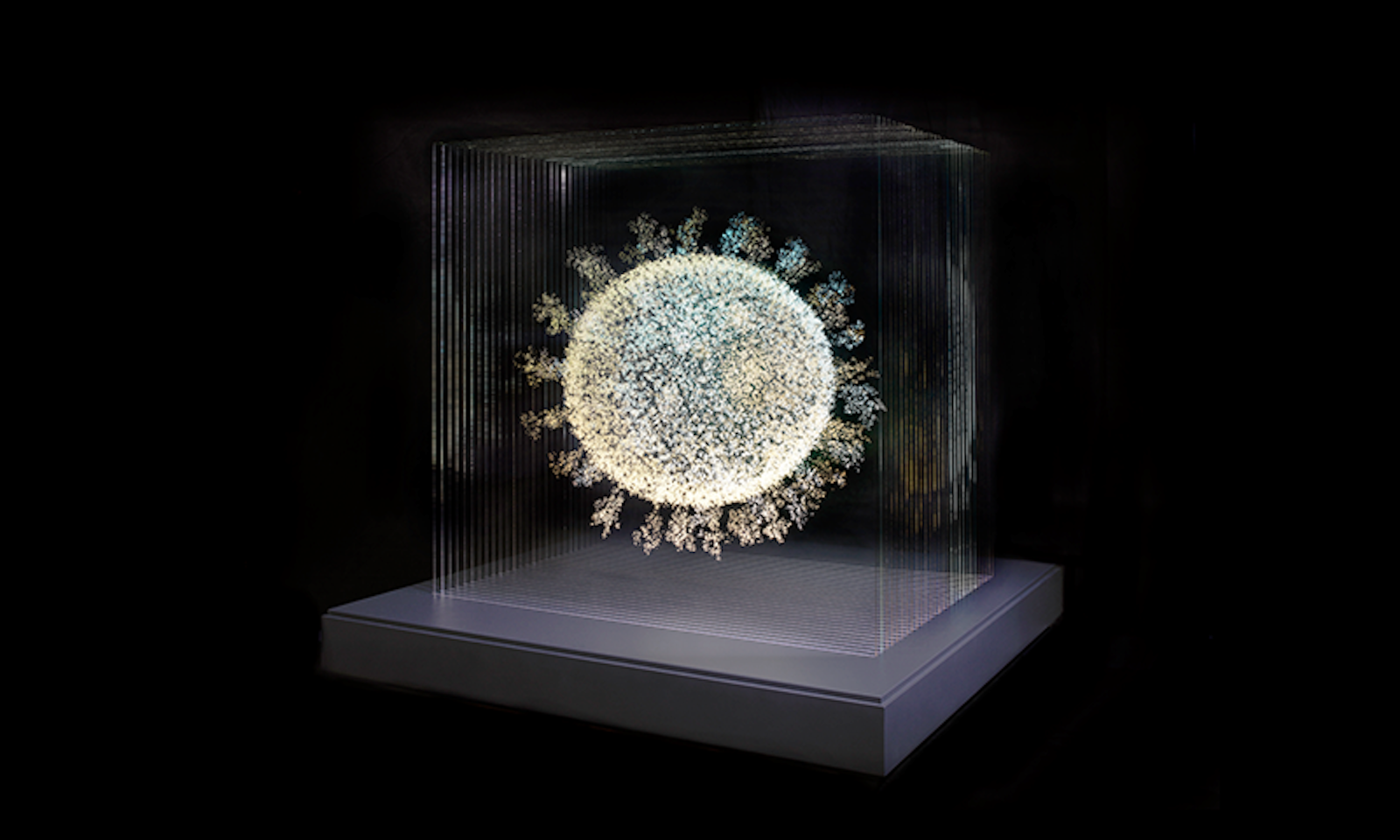
First exhibited at the Oxford Museum of Natural History, 2020: The Sphere That Changed the World, is a scrupulously accurate three-dimensional “drawing” of the virus by Scottish artist Angela Palmer. It appears to have trapped a single SARS-CoV-2 viral particle in glass, magnified to 8 million times its size. The effect is chilling. The sphere appears otherworldly, suspended in time and space, lit from within with a dizzying clarity.
The shape of Palmer’s work is based on the precise modeling of the virus using bioinformatic techniques and the computational outputs of one of the world’s most powerful supercomputers, located at the Texas Advanced Computing Center in Austin. Using the genetic code of the first isolated strain of SARS-CoV-2, scientists from the United States and Netherlands were able to predict the arrangement of proteins and lipids that make up the outer surface of the virus. Palmer fed this information into industrial design software and using a technique of hand-engraving transferred cross-sections of the outer shell onto 28 sheets of glass stacked together to produce the 3-D result.
After studying anatomy at Oxford University’s Ruskin School of Drawing and Fine Art, Palmer “became fascinated in visually representing internal structures hidden beneath the surface,” she wrote to me in an email correspondence. “I like to peel back the layers and expose the beauty of natural forms. During lockdown, I became determined to create the one form which was dominating humankind. Its invisibility made it a natural subject for me.”

Palmer sought out scientists who could provide a blueprint of the virus, which she would use to create her sculpture. In earlier work, the artist had used MRI and CT scan images to produce anatomical works to great success, including a “self-portrait” of her brain, exhibited at the National Portrait Gallery of Scotland. But finding accurate tomographic data that informed the shape of the virus proved harder than she expected.
When it comes to visualization, viruses fall into the liminal space between cell and molecule. We have powerful techniques that can probe three-dimensional structures at each side of this spectrum but are hindered by a blind spot in between. A single SARS-CoV-2 particle is one thousand times smaller than the width of a human hair. There are no magnifying lenses that are powerful enough to bring it into view, and even our most probing cryo-electron microscopy techniques, which can be used to study the anatomy of a cell, fall short on resolution. On the smaller side of the size spectrum, techniques such as X-ray crystallography and NMR spectroscopy can provide insight into the shape of molecules such as DNA and proteins, but from this perspective the viral particles are too large to comprehend. Just like Alice in Wonderland, we seem to struggle to bring ourselves into the right size frame for perspective.
But scientists went through nature’s looking glass to uncover another means of visualization—the genetic code that a virus uses to assemble itself. If this code can be correctly interpreted, it is possible to conjure the virus’ physical structure inside the brain of a supercomputer. “This is precisely where sci-fi becomes reality—because of [structural] bioinformatics,” Dmitry Korkin, a professor in the bioinformatics and computational biology program at Worcester Polytechnic Institute, told me. Early in 2020, his team produced “the periodic table of structural elements that comprise the SARS-CoV-2 virus,” based on the genetic sequence information of the first isolated strain from Wuhan, China.
The artwork’s accuracy offers a key to the virus’s undoing.
The outbreak of SARS-CoV-2 sent the scientific community into a frenzy of work and collaboration. “We worked in a hackathon. We had meetings at 3 a.m., pushing to characterize the virus,” Korkin said. Rather than wait for the formal process of peer review and publication, his group released all findings directly onto their website. Traditional news outlets broadcasted the results, teenagers in China spread the infographics on TikTok, and scientists reached out to consolidate their knowledge.
The clinching moment for producing a plausible 3-D model of the virus came after a fruitful collaboration with Siewert-Jan Marrink’s group at the University of Groningen in the Netherlands. Marrink studies the dynamics of what to Korkin’s eyes are “very large systems”—the lipid membranes of cells. But the frame for the SARS-CoV-2 sphere happens to be a lipid bilayer that the virus steals from the cells of its host.
Together the scientists authored a successful grant petitioning for time on a supercomputer at the Texas center, where they could model all the components of the viral sheath: the proteins embedded in the lipid bilayer which coalesce to form its outer surface. The pieces of the puzzle were known, all that remained was to determine a structure that did not break any physical laws of nature. The genetic code predicts the individual proteins, but it is not clear how many copies of each protein together make a single sphere, in what proportion or configuration.
The scientists tasked the supercomputer to produce iterative simulations of various combinations of the known viral proteins behaving in three-dimensional space over the course of hundreds, up to a thousand nanoseconds. Each iteration, where each nanosecond takes the supercomputer approximately a day of computation, is just long enough to see whether the structure undergoes any “catastrophic events,” according to Korkin. “If you introduce the wrong number of ions”—charged particles—“into the simulation, the virus’ shell will explode, or if you introduce the wrong proportion of lipids, or if you try to squeeze in too many proteins, it is going to rupture.”
It took a week on the computing server to produce a reliable simulation, and then a few more to test the model to perfection. The result was an imperfect sphere, slightly flattened at the top and bottom, with membrane, spike, and envelope proteins coalescing together in the lipid bilayer in stable proportions. This was the model shared with Palmer, dissected into 28 cross-sections.
Palmer’s scientifically accurate representation of the SARS-CoV-2 virus is not strictly symbolic. Nor is it a simple unmasking of the enemy, a way to give body and form to our collective fear. The accuracy of the artwork provides a map and key to the virus’ undoing. Knowing what a virus looks like helps scientists understand its mode of attack, notably where its structure interlocks with the receptors on cells. It foreshadows where a virus could be most vulnerable to counterattack by targeted drugs.
“When I finished, I was taken aback by its beauty.”
When describing the exciting discoveries from the modeling effort, Korkin referred to the membrane proteins as revealing themselves to be the “hidden heroes of the virus.” Simulations of their behavior showed them assembling into filaments folding into a mesh structure, providing a flexible but durable reinforcement of the membrane sheath. Equipped with this knowledge, researchers targeted the chinks in SARS-CoV-2’s armor.
One way that Palmer’s work is limited in its depiction is that, as any solid sculpture, it represents the viral body as rigid, locked into shape. “This virus is everything but that,” Korkin explained to me. With a lipid frame it can pulsate, squeeze, and maneuver its way around the cells in our body.” His own simulations, Korkin said, produced in his mind’s eye the fact that the “outer shell almost breathes.” Palmer’s chosen medium of glass also gives the virus a false appearance of fragility. Here is a particle that is extremely durable, highly resistant to physical wear and tear, malleable and adaptive.
Palmer admits that her sculpture is full of paradox and allusion. It traps a virus that has in turn held all of us captive over the course of a pandemic. And while it is rendered in solid form, she likes to emphasize its elusiveness: “As you confront the sculpture head on, you see the entire sphere, with its protein spikes emerging from its surface. But as you move around it, past the stacked glass sheets, the virus particle disappears entirely from view, only to reappear again, just as it torments us.” Perhaps its greatest paradox is its visual appeal. It is a shimmering ode to a killer agent that has taken the lives of millions around the globe.
“When I finally finished the engravings and stacked up the glass to form the entire sphere, I was taken aback by its beauty,” Palmer said. “As an artist representing a deadly object, which is destroying lives and livelihoods worldwide—described by AstraZeneca pioneer Sarah Gilbert as our ‘mortal enemy’—you might expect to create something that is menacing and frightening. But in reality it totally contradicted that. I found it immediately beautiful and mesmerizing. It’s a natural form, and I can’t think of any natural forms which are not beautiful.”
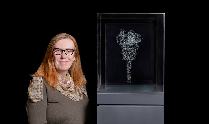
2020: The Sphere That Changed the World was acquired by the Science Museum in London, where it is exhibited as part of the permanent collection. 
Elena Kazamia is a scientist and freelance journalist with a Ph.D. in plant sciences from the University of Cambridge. She is originally from Greece.
Lead photo courtesy of Andrew Smart of A.C.Cooper Ltd